Acute megakaryoblastic leukemia
| Acute megakaryoblastic leukemia | |
|---|---|
|
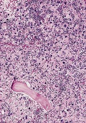
AML-M7, bone marrow section | |
| Classification and external resources | |
| Specialty | Hematology and oncology |
| ICD-10 | C94.2 |
| ICD-O | M9910/3 |
| MeSH | D007947 |
Acute megakaryoblastic leukemia (AMKL) is a form of leukemia where a majority of the blasts are megakaryoblastic.[1]
It is classified under AML-M7 category of the French-American-British classification.[2]
The latest WHO classification (2008, Lyon), classifies Acute Myeloid Leukemia into distinct subtypes, based on clinico-pathological and molecular features. Acute megakaryoblastic leukemia is placed under the AML-Not Otherwise Specified subcategory.
Diagnosis requires more than 20% Blasts in the marrow/ peripheral blood with more than 50% demonstrating megakaryocytic derivation by morphology, immunophenotypic or electron microscopic studies.
Causes
It is associated with GATA1, and risks are increased in individuals with Down syndrome.[3]
However, not all cases are associated with Down syndrome,[4] and other genes can also be associated with AMKL.[5]
Another related gene is MKL1, which is also known as "MAL".[6] This gene is a cofactor of serum response factor.[7]
Signs and symptoms
In adults, include pancytopenia with low blast counts in the blood, myelofibrosis, an absence of lymphadenopathy and hepatosplenomegaly, poor response to chemotherapy, and short clinical course. In children, the same clinical presentation but with variable course especially in very young children; both leukocytosis and organomegaly may be present in children with M7.
In the first three years of life, megakaryoblastic leukemia is the most common type of leukemia in patients with Down syndrome.[8]
Diagnosis
The morphology of cells was observed by means of bone marrow smear; the immunophenotype was detected by flow cytometry and immunohistochemistry assay.[9]
Blasts more than 20%, with more than 50% of megakaryocytic phenotype.
In blood and bone marrow smears megakaryoblasts are usually medium-sized to large cells with a high nuclear-cytoplasmic ratio. Nuclear chromatin is dense and homogeneous. There is scanty, variable basophilic cytoplasm which may be vacuolated. An irregular cytoplasmic border is often noted in some of the megakaryoblasts and occasionally projections resembling budding atypical platelets are present. Megakaryoblasts lack myeloperoxidase (MPO) activity and stain negatively with Sudan black B. They are alpha naphthyl butyrate esterase negative and manifest variable alpha naphthyl acetate esterase activity usually in scattered clumps or granules in the cytoplasm. PAS staining also varies from negative to focal or granular positivity, to strongly positive staining. A marrow aspirate is difficult to obtain in many cases because of variable degree of myelofibrosis. More precise identification is by immunophenotyping or with electron microscopy (EM). Immunophenotyping using MoAb to megakaryocyte restricted antigen (CD41 and CD61) may be diagnostic.[10]
Prognosis
Complete remission and long-term survival are more common in children than adults.
Prognosis depends upon the cause. One third of cases is associated with a t(1;22)(p13;q13) mutation in children. These cases carry a poor prognosis.[11]
Another third of cases is found in Down syndrome. These cases have a reasonably fair prognosis.
The last third of cases may be heterogeneous, and carry a poor prognosis.[12]
See also
References
- ↑ "Final Diagnosis -- Case 439". Archived from the original on 24 February 2008. Retrieved 2008-03-08.
- ↑ "Acute Myeloid Leukemia - Signs and Symptoms".
- ↑ Hitzler JK, Cheung J, Li Y, Scherer SW, Zipursky A (2003). "GATA1 mutations in transient leukemia and acute megakaryoblastic leukemia of Down syndrome". Blood. 101 (11): 4301–4. doi:10.1182/blood-2003-01-0013. PMID 12586620.
- ↑ Hama A, Yagasaki H, Takahashi Y, et al. (2008). "Acute megakaryoblastic leukaemia (AMKL) in children: a comparison of AMKL with and without Down syndrome". Br. J. Haematol. 140 (5): 552–61. doi:10.1111/j.1365-2141.2007.06971.x. PMID 18275433.
- ↑ Gu TL, Mercher T, Tyner JW, et al. (2007). "A novel fusion of RBM6 to CSF1R in acute megakaryoblastic leukemia". Blood. 110 (1): 323–33. doi:10.1182/blood-2006-10-052282. PMC 1896120
 . PMID 17360941.
 - ↑ Mercher T, Coniat MB, Monni R, et al. (May 2001). "Involvement of a human gene related to the Drosophila spen gene in the recurrent t(1;22) translocation of acute megakaryocytic leukemia". Proc. Natl. Acad. Sci. U.S.A. 98 (10): 5776–9. doi:10.1073/pnas.101001498. PMC 33289
 . PMID 11344311.
 - ↑ Vartiainen MK, Guettler S, Larijani B, Treisman R (June 2007). "Nuclear actin regulates dynamic subcellular localization and activity of the SRF cofactor MAL". Science. 316 (5832): 1749–52. doi:10.1126/science.1141084. PMID 17588931.
- ↑ Hitzler, JK (2007). "Acute megakaryoblastic leukemia in Down syndrome". Pediatric blood & cancer. 49 (7 Suppl): 1066–9. doi:10.1002/pbc.21353. PMID 17943965.
- ↑ Lei Q, Liu Y, Tang SQ (2007). "[Childhood acute megakaryoblastic leukemia]". Zhongguo Shi Yan Xue Ye Xue Za Zhi (in Chinese). 15 (3): 528–32. PMID 17605859.
- ↑ Vardiman JW, Harris NL, Brunning RD (2002). "The World Health Organization (WHO) classification of the myeloid neoplasms". Blood. 100 (7): 2292–302. doi:10.1182/blood-2002-04-1199. PMID 12239137.
- ↑ Huret JL . t(1;22)(p13;q13) Archived July 3, 2008, at the Wayback Machine.. Atlas Genet Cytogenet Oncol Haematol.
- ↑ Cuneo A, Cavazzini F, Castoldi GL . Acute megakaryoblastic leukemia (AMegL), M7 acute non lymphocytic leukemia (M7-ANLL). Atlas Genet Cytogenet Oncol Haematol.
