Acoustic tweezers
Acoustic tweezers is a technology that is able to control the movement of objects by sound waves. In a standing acoustic field, objects will experience an acoustic radiation force that moves the objects to special regions of the acoustic field.[1] Depending on the properties (density, compressibility) of the objects, they can be moved to either acoustic pressure nodes (minimum pressure regions) or pressure antinodes (maximum pressure regions).[2] As a result, precise manipulation of objects using sound waves is feasible by controlling the position of pressure nodes. Acoustic tweezers does not require expensive equipment and complex experimental setups. More importantly, acoustic waves have been proven safe to biological objects, making it an ideal tool for biomedical applications.[3] In recent years, acoustic tweezers has found many important applications in the area of manipulating sub-millimeter objects, such as flow cytometry, cell separation, cell trapping, single-cell manipulation, and nanomaterial manipulation.[4] The use of one-dimensional standing waves to manipulate small particles was reported for the first time in "Ultrasonic inspection of fiber suspensions".[5]
Fundamental theory
Particles in acoustic field will be moved by forces originated from the interaction among the acoustic waves, fluid and the particles. These forces, including acoustic radiation force, secondary field force between particles, and Stokes drag force, create the phenomena of acoustophoresis, which is the foundation of acoustic tweezers technology.
Acoustic radiation force
When a particle is suspended in the field of a sound wave, a so-called acoustic radiation force, which arises from the scattering of the acoustic waves on the particle, will exert on the particle. The studies of acoustic radiation forces on suspended particles have a long history. The force was first modelled and analyzed for incompressible particles in an ideal fluid by King in 1934.[6] Yosioka and Kawasima calculated the acoustic radiation force on compressible particles in a plane wave field in 1955.[7] Gorkov summarized the previous work and proposed equations to determine the average force acting on a particle in an arbitrary acoustical field when its size is much smaller than the wavelength of the sound.[1] Recently, Bruus revisited the problem and gave detailed derivation for the acoustic radiation force.[8]

As shown in Figure 1, the acoustic radiation force on a small particle results from a non-uniform flux of momentum in the near-field region around the particle, which is caused by the incoming acoustic waves and the scattering on the surface of the particle when acoustic waves propagate through it. For a compressible spherical particle with a diameter much smaller than the wavelength of acoustic waves in an ideal fluid, the acoustic radiation force can be calculated by , where is a given quantity, also called acoustic potential energy.[1][8] The acoustic potential energy is expressed as:
where
• is the particle volume,
• is the acoustic pressure,
• is the velocity of acoustic particles,
• is the fluid mass density,
• is the speed of sound of the fluid,
• , are two coefficients.
• is the time-average term,
The two coefficients and can be calculated by ,
where
• is the mass density of the particle,
• is the speed of sound of the particle.
Acoustic radiation force in standing waves
The standing waves can form stable acoustic potential energy field, so they are able to create stable acoustic radiation force distribution, which is desirable for many acoustic tweezers applications. For 1-D planar standing waves, the acoustic fields are given by:[8]
,
,
,
where
• is the displacement of acoustic particle,
• is the acoustic pressure amplitude,
• is the angular velocity,
• is the wave number.
With these fields, the time-average terms can be obtained, and they are:
,
,
Thus, the acoustic potential energy is:
,
Then, the acoustic radiation force is found by differentiation:
,
, ,

where is the acoustic energy density, and is acoustophoretic contrast factor. The term shows that the radiation force period is one half of the pressure period. Also, the contrast factor can be positive or negative depending on the properties of particles and fluid. For positive value of , the radiation force points from the pressure antinodes to the pressure nodes, as shown in Figure 2, and the particles will be pushed to the pressure nodes.
Secondary acoustic forces
When multiple particles in a suspension are exposed to a standing wave field, they will not only experience acoustic radiation force, but also secondary acoustic forces caused by waves scattered by particles. The interparticle forces are sometimes called Bjerknes forces. A simplified equation for the interparticle forces:[9][10]
where
• is the radius of the particle,
• is the distance between the particles,
• is the angle between the central line of the particles and the direction of propagation of the incident acoustic wave.
The sign of the force represents the direction of the force. A negative sign means an attractive interparticle force, whereas a positive sign means a repulsive force. The left side of the equation depends on the acoustic particle velocity amplitude and the right side depends on the acoustic pressure amplitude . When particles are lined up in the direction of the acoustic wave propagation (θ=0˚) the velocity-dependent term is repulsive, and likewise attractive when the particles are perpendicular (θ=90˚) to the incident wave propagation. The pressure-dependent term is not affected by the particle orientation at all and is always attractive. In the case of positive contrast factor, the velocity-dependent term diminishes as particles are driven to the velocity node (pressure antinode), as in the case of air bubbles and lipid vesicles. In a similar way the pressure-dependent term diminishes as particles are driven towards the pressure node (velocity antinode), as are most solid particles in aqueous solutions. The influence of the secondary forces is usually very weak, due to the distance term d in the denominator, which means that it is only effective when the distance between particles is very small. The secondary force becomes important in aggregation and sedimentation applications, where particles initially are gathered in nodes by the acoustic radiation force, and as interparticle distances become smaller the secondary forces assist in a further aggregation until the clusters finally become heavy enough for the gravity to overcome the buoyancy and start the sedimentation process.

Acoustic streaming
Acoustic streaming is a steady flow generated by a nonlinear effect in an acoustic field. Depending on the mechanisms, the acoustic streaming can be categorized into two general types, Eckert streaming and Rayleigh streaming.[11][12] Eckert streaming is driven by a time-average momentum flux created when high amplitude acoustic wave propagates and attenuates in fluid. Rayleigh streaming, also called “boundary driven streaming”, is forced by a shear viscosity close to a solid boundary. Both of the driven mechanisms come from a time-average nonlinear effect. Regarding to the nonlinearity of acoustic streaming, a so-called perturbation approach is used to analyze this phenomena.[13] The governing equations for this problem are mass conservation and Navier-Stokes equations:
,
where
• is the density of fluid,
• is the velocity of fluid particle,
• is the pressure
• is the dynamic viscosity of fluid
• is the viscosity ratio.
The perturbation series can be written as , , ,where the higher order terms are much smaller than the lower order ones.
The liquid is quiescent and homogeneous at its zero-order state. Substituting the perturbation series into the mass conservation and Navier-Stokes equation and using the relation of , the first-order equations can be obtained by collecting terms in first-order,
,
.
Similarly, the second-order equations can be found as well,
,
.
For the first-order equations, if take the time derivation of the Navier-Stokes equation and insert the mass conservation, a combined equation can be found,
.
This equation is actually an acoustic wave equation with viscous attenuation. Physically, and can be interpreted as the acoustic pressure and the velocity of acoustic particle, respectively.
For the second-order equations, they can be considered as governing equations using to describe the motion of fluid with mass source and force source . Generally, the acoustic streaming is a steady mean flow, whose response time scale is much smaller than the one of the acoustic vibration. The time-average term is normally used to present the acoustic streaming. By time average the second-order equations and using that , the time-average second-order equations can be obtained,
,
.
To get the acoustic streaming, the first-order equations should be solved first. Since Navier-Stokes equations can only be solved for some simple cases analytically, numerical methods are the most used option to work them out in engineering. Finite element method (FEM) is one of the most used numerical method. It can be employed to simulate the acoustic streaming phenomena. Figure 3 is one example of acoustic streaming around a solid circular pillar, which is calculated by FEM method.
As said, the acoustic streaming is driven by mass and force sources originating from the acoustic attenuation. However, they are not the only driven forces for the acoustic streaming. The boundary vibration may also have contribution to the acoustic streaming, especially to “boundary driven streaming”. For these cases, the boundary condition should also be processed by perturbation approach and be imposed to the two order equations accordingly.
Particle motion
The motion of a suspended particle whose gravity is balanced by the buoyance force in an acoustic field is determined by two forces: the acoustic radiation force and Stokes drag force. By applying the Newton’s law, the motion can be described as:
,
.
where
• is the fluid velocity,
• is the velocity of particle.
For applications in a static flow, the fluid velocity comes from the acoustic streaming. The magnitude of acoustic streaming depends on the power and frequency of input. Also, the properties of fluid media affect the value as well. For typical acoustic-based microdevices, the operating frequency ranges from ~kHz to ~MHz. The vibration amplitude is in range of 0.1 nm~1 µm. The fluid used is water. The estimated magnitude of acoustic streaming is in range of 1 µm/s~ 1 mm/s. Thus, the acoustic streaming should be smaller than the main flow for most continuous flow applications. The drag force is mainly induced by the main flow in those applications.
Applications
Label-free cell separation
As an example, acoustic tweezers can be used in separation of lipid particles from erythrocytes. During cardiac surgery supported by a heart-lung machine a massive embolization of lipid particles occurs in the brain when shed blood is returned to a patient via a filter. The lipid particles are derived from triglycerides leaking from fat cells during surgery in adipose tissue. The embolization is associated with cognitive dysfunction observed after surgery. The techniques currently available for blood wash do not meet the demand to remove these lipid particles. The most common method to wash blood is based on centrifuges which are burdened with a number of drawbacks, i.e. they only handle larger amounts of blood (#0.5 l), are harmful for the blood cells, need specially trained personnel, are not continuous and display a limited lipid particle elimination.

The technology presented in this communication offers a solution to the embolization problem by employing the possibility of discriminating erythrocytes from lipid particles. In addition, when fully developed and implemented clinically, it reduces the demand for allogenic blood and reduces or eliminates blood transfusion related incompatibilities. The primary acoustic radiation force equation tells us that the acoustic force can move particles either towards a node or an anti-node of a standing wave depending on their densities and compressibilities. If the particles are red blood cells and lipid droplets in blood plasma, the erythrocytes gather in the pressure node (in the centre of the channel) while the lipid particles gather in the pressure anti-nodes (by the side walls), Fig. 4. At the end of the channel the red blood cells exit through the centre outlet while the lipid particles exit through the side outlets, separating the two particle types, Fig. 5. visual observations of the separation process performed on human blood could be made, displaying steady streams of lipid particles flowing along the side walls and a well defined band of red blood cells exiting the system through the centre outlet channel, Fig. 6.

In addition to separate particles with different sign of acoustic contrast factors, acoustic method can also be used to separate particles with different size. Shi et al. reported a standing surface acoustic wave (SSAW) microfluidic device to separate microparticles with different diameters.(Fig. 7)[14] According to the equation of primary acoustic radiation force, larger particles with experience larger force than smaller particles. Shi et al. used interdigital transducers (IDTs) to generate SSAW field in a microfluidic channel, and they positioned the pressured nodes in the middle of the channel. When introducing a mixture of particles with different size from the edge of the channel, larger particles will migrate faster to middle of the channel and be collected at the center outlet. Smaller particles will not be able to migrate to the center outlet before they are collected from the side outlets. This experiment setup has also been used to separate blood components, bacteria, and hydrogel particles.[15][16][17]

Fluorescence-activated cell sorting
Fluorescence-activated cell sorters (FACS) are powerful tools for high-throughput single-cell characterization and sorting. A typical FACS system includes three major modules: (1) a fluidic module to three-dimensionally focus the stream of biological cells, (2) an optical module to detect fluorescence emissions and scattered light arising from individual cells, and (3) a cell-sorting module to separate cells of interest from other cells. Although FACS has a variety of applications in basic biomedical research, clinical practice, and clinical trials, current benchtop FACS systems have the following drawbacks: high cost, large size, complex configuration, and high maintenance costs. In addition to these well-known draw backs, the biocompatibility of conventional FACS system has always been a practical concern for the users. In a conventional FACS, there are three major factors affecting cell normal physiology: (1) High shear pressure that cells experience during the cell-focusing process; (2) High voltage and electric field needed during the cell-sorting process; (3) High impact forces that cells experience during the cell-sorting process. These harsh conditions may result in the immediate cell damage and the change of gene expression. In the past decade, the application of acoustic tweezers toward improving the design of FACS has shown great potential to overcome these limitations. By integrating the acoustic tweezers with optical/electrical modules, a miniaturized microfluidic platform is established for simultaneous cell analysis and sorting, which are safe to cells. With the advantages of low cost, small size, high biocompatibility, and high biosafety, acoustic tweezers enabled FACS system will not only be an excellent replacement for the benchtop counterparts, but also fulfill many unmet needs in biomedical studies and clinical applications, such as the applications involving sensitive or fragile cells, low-volume or low-abundance samples.
Acoustic tweezers have been developed to achieve 3D focusing of cells/particles in microfluidics. A schematic of the standing surface acoustic wave (SSAW) focusing device is shown in Fig. 5.[18] A pair of interdigital transducers (IDTs) are deposited on a piezoelectric substrate, and a microfluidic channel is bonded with the substrate and positioned between the two IDTs. Microparticle solutions are infused into the microfluidic channel by a pressure-driven flow. Once an RF signal is applied to both IDTs, two series of surface acoustic waves (SAW) propagate in opposite directions toward the particle suspension solution inside the microchannel. The constructive interference of the two SAWs results in the formation of a SSAW, as well as the periodic distribution of the pressure nodes (minimum pressure amplitude) and anti-nodes (maximum pressure amplitude) on the substrate. When the SSAW encounter the liquid medium inside the channel, leakage waves in the longitudinal mode are generated, causing pressure fluctuations in the medium. These pressure fluctuations result in acoustic radiation forces that act laterally (in the x-direction of Fig. 8) on the particles. As a result, the suspended particles inside the channel will be forced toward either the pressure nodes or antinodes, depending on the density and compressibility of the particles and the medium. When the channel width covers only one pressure node (or antinode), the particles will be trapped in that node and consequently, focusing is achieved.

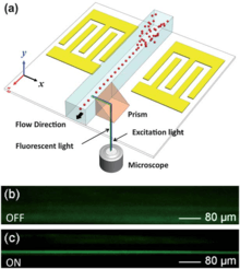
Fig. 9 shows the focusing of polystyrene particles (top) and HL-60 cells (bottom) from top view using acoustic tweezers. In addition to focusing in horizontal direction, cells/particles are also focused in the vertical direction with acoustic tweezers as shown in Fig. 10.[19] To observe the side view, a 45-degree prism adjacent to the channel is used to bend the excitation/fluorescent light for observation of particle migration in the vertical direction. After SSAW is on, the randomly distributed particles (Fig. 10b) are focused into a single file stream (Fig. 10c) in the vertical direction. By integrating a standing surface acoustic wave (SSAW)-based microdevice capable of 3D particle/cell focusing with a laser-induced fluorescence (LIF) detection system, acoustic tweezers are developed into a microflow cytometer for high-throughput single cell analysis.


Fig. 11A shows a typical data record of the fluorescent peaks using calibration beads.[20] The uniform peak intensities in the data plot indicate a consistent measurement of the calibration particles. The peak intensities exhibit a Gaussian distribution as shown in Fig. 11B. The fluorescent signals without SSAW focusing are shown in Fig. 11C as a negative control for comparison. Because the non-focused particles have different trajectories in the microchannel, a large number of them miss the excitation light spot or moved in different planes along the vertical direction, resulting in a much lower number of peaks detected with a larger non-uniformity in peak intensities. The SSAW-based 3D focusing enabled microfluidic cytometer does not require any sheath flows or complex structures, and it allows for simple operation over a wide range of sample flow rates. Moreover, with the gentle, bio-compatible nature of low-power surface acoustic waves, the acoustic tweezers is expected to be able to preserve the integrity of cells and other bioparticles.

In addition to 3D cell focusing, acoustic tweezers can also be used as a unit for cell sorting. A schematic of SSAW-based, on-chip, multichannel cell-sorting technique is shown in Fig. 12, which takes advantages of the tunability offered by chirped interdigital transducers (IDTs).[21][22] The SSAW-based sorting method manipulates the objects (i.e., cells, particles, and droplets) directly by the acoustic radiation force with excellent controllability and a large range of translation, which renders it capable of precisely sorting cells into a great number (e.g., five) of outlet channels in a single step. This is a major advantage over most existing sorting methods, which typically only sort cells into two outlet channels. This approach is simple, versatile, and compact, that can be valuable in many chemical or biological analytical processes to facilitate the development of micro total analysis systems (μTAS).

Noninvasive cell trapping and patterning
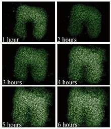
Noncontact trapping and retention of cells in microfluidic networks by means of acoustic standing wave forces are demonstrated as a platform for perfusion-based cell handling and assaying. The current improvements of the described acoustic trapping microfluidic platform provide stable resonator dimensions over extended periods of operation. A key feature is the manufacturing of the microfluidic channels directly in the glass reflector layer, thus avoiding the use of photolithography-defined polymer gaskets that may undergo swelling during the experiments. This now enables longer periods of stable operation of the noncontact acoustic particle/cell trapping platform as demonstrated in the cell culturing experiment over 6 h. The temperature measurements indicate that the thermal environment in the acoustic trap is at a level where no negative effects are expected on cell behavior. The viability tests verify that the acoustic intensities used give no indication of being harmful to the cells, but this will have to be confirmed by extended studies, including gene expression profile analysis.
Figure 13. (a) Side-view schematic of the microfluidic device. The glass reflector with etched fluidic channels is clamped to the PCB holding the transducer. Cells infused into the chip are trapped in the ultrasonic standing wave formed in the channel. The acoustic forces focus the cells into clusters in the center of the channel as illustrated in the inset. (b) Since the trapping occurs close to the transducer surface, the actual trapping sites are given by the near-field pressure distribution as shown in the 3D image. Cells will be trapped in clusters around the local pressure minima creating different patterns depending on the amount of cells trapped. The peaks in the graph correspond to the pressure minima.

Figure 14. Growth of YFP-expressing yeast cells, UMR106, trapped in the acoustic device while being perfused with cell medium. The images show the increase of the number of cells in the cell cluster after 1-6 h of cultivation. The successful growth indicates that the cell proliferation is not affected by the high-frequency acoustic radiation. The horseshoe-shaped pattern is caused by the cells clustering in the near-field pattern.

Fig. 15A shows the schematic of a standing surface acoustic waves (SSAW)-based cell enrichment device.[23] A microchannel is assembled in the SSAW-activated region of the substrate with its long axis oriented in the propagation direction of acoustic waves. In order for the acoustic waves to propagate into the microchannel, a coupling gel is used to fill the gap between the tubing and the substrate, as shown in Fig. 15B. Applying an AC signal to the IDTs results in the generation of a SSAW field and thus the creation of a non-uniform pressure field in the fluid with a periodic distribution of pressure nodes and antinodes. In the presence of the SSAW field, a cell suspension is injected into the microchannel. Upon entering the region where the coupling gel bonds the microchannel to the substrate, known as the enrichment region, cells are trapped at SSAW pressure nodes. As more fluid pass through the enrichment region, the concentration of the trapped cells gradually increase. Finally, the SSAW is turned off to release the cells. A parallel sample enrichment approach could help to improve the working efficiency and throughput by enriching more cells in less time, or enriching different species of cells simultaneously. As shown in Fig. 15D, by assembling an array of microchannels onto the piezoelectric substrate, parallel sample enrichment is conveniently established. In each channel, the efficiency of the sample enrichment is consistent (Fig. 15E), revealing a steady performance of each working unit.

As shown in Fig. 16,[24] acoustic tweezers can be used as an active patterning technique that utilizes standing surface acoustic wave (SSAW) to manipulate and pattern cells and microparticles. This technique is capable of patterning cells and microparticles regardless of shape, size, charge or polarity. Its power intensity, approximately 500,000 times lower than that of optical tweezers, compares favorably with those of other active patterning methods. Flow cytometry studies have revealed it to be non-invasive. The aforementioned advantages, along with this technique’s simple design and ability to be miniaturized, render the ‘‘acoustic tweezers’’ technique a promising tool for various applications in biology, chemistry, engineering, and materials science.

Fig. 17 shows acoustic-based tunable patterning technique by which microparticles or cells can be arranged into reconfigurable patterns in microfluidic channels.[25] In this approach, two pairs of slanted-finger interdigital transducers (SFITs) are to generate a tunable standing surface acoustic wave field, which in turn patterns microparticles or cells in one- or two-dimensional arrays inside the microfluidic channels—all without the assistance of fluidic flow. By tuning the frequency of the input signal applied to the SFITs, the cell pattern can be controlled with tunability of up to 72%. This acoustic-based tunable patterning technique has the advantages of wide tunability, non-invasiveness, and ease of integration to lab-on-a-chip systems, and shall be valuable in many biological and colloidal studies.

Manipulation of single cell, particle, or organism
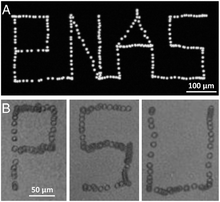
Manipulating single cells is important to many biological studies, such as in controlling the cellular microenvironment and isolating specific cells of interest. Acoustic tweezers has been demonstrated to manipulate each individual cell with micrometer-level resolution. Cells generally have a diameter of 10–20 μm. To meet the resolution requirements of manipulating single cells, short-wavelength acoustic waves should be employed. In this case, surface acoustic wave (SAW) is a more favored option than bulk acoustic wave (BAW) as it allows using shorter-wavelength acoustic waves (normally less than 200 μm).[26] Ding et al. reported a surface standing acoustic wave (SSAW) microdevice that is able to manipulate single cells with prescribed paths.[27] Fig.18 shows that the movement traces of single cells can be controlled to form the letter sequences PNAS and PSU, respectively. The working principle of the device lies in the controlled movement of pressure nodes in a SSAW field. Ding et al. employed chirped interdigital transducers (IDTs) that are able to generate SSAWs with adjustable positions of pressure nodes. By simply changing the input frequency, the position of pressure nodes can be changed. Ding et al. also showed that not only micrometer-sized cells, but also the millimeter-sized microorganism C. elegan can be manipulated with the same manner. In this manner, it can be programmed to move single cells along any desired paths. It should be noted that Ding et al. also examined cell metabolism and proliferation after the acoustic treatment. No significant differences were found comparing to the control group, indicating the non-invasive nature of acoustic base manipulation. In addition to using chirped IDTs, phase shift based single particle/cell manipulation has also been reported.[28][29][30]

Manipulation of single (bio)molecules
Recently, Sitters et al.[31] have shown that acoustics can be used to manipulate single (bio) molecules such as DNA and proteins. This method, which the inventors name Acoustic Force Spectroscopy, allows one to measure the force response of single molecules. This is achieved by attaching small micro spheres to the molecules at one side and attaching them to a surface at the other. By pushing the micro spheres away from the surface with a standing acoustic wave the molecules are effectively stretched out.
Manipulation of organic nanomaterials
In recent years, polymer-dispersed liquid crystals (PDLCs) have been used in many applications including smart windows, displays, microlenses, lasers, and data storage, due to their excellent electro-optical properties. Within a PDLC film, liquid crystals (LCs) are generally trapped in a transparent polymer medium, thus forming micrometer-scale LC droplets. The PDLC film can be switched from opaque to transparent by acoustic tweezers. A SAW-driven PDLC light shutter has been demonstrated by integrating a cured PDLC film and a pair of interdigital transducers (IDTs) onto a piezoelectric substrate.[32] The IDTs were used to generate the SAW for driving the PDLC film. Figure 19 illustrates the layout and working principle of the SAW-driven PDLC light shutter. A PDLC cell is located in between two identical IDTs, which are deposited on a piezoelectric substrate with a parallel arrangement. One IDT is used for SAW generation and the other is used for SAW detection. A radio-frequency (RF) signal is applied to a single IDT to generate a SAW, which propagates along the surface of the piezoelectric substrate (x direction in Figure 19). By tuning the applied frequencies from the function generator and monitoring the frequency-dependent output voltage from the detection IDT that is connected to an oscilloscope, an optimal resonant frequency can be selected for the driving frequency. Under this resonance frequency, the LC molecules can be reoriented perpendicular to the substrate. Therefore, the optical properties of PDLC film can be changed with acoustic tweezers.

Manipulation of inorganic nanomaterials
Patterning of nanowires in a controllable, tunable manner is important for the fabrication of functional nanodevices. Acoustic tweezers provide a simple approach for tunable nanowire patterning (Fig. 20).[33] This technique allows for the construction of large-scale nanowire arrays with well-controlled patterning geometry and spacing within 5 s. In this approach, SSAWs are generated by interdigital transducers, which induced a periodic alternating current (AC) electric field on the piezoelectric substrate and consequently patterned metallic nanowires in suspension. The patterns could be deposited onto the substrate after the liquid evaporated. By controlling the distribution of the SSAW field, metallic nanowires are assembled into different patterns including parallel and perpendicular arrays. The spacing of the nanowire arrays could be tuned by controlling the frequency of the surface acoustic waves. Additionally, 3D spark-shaped nanowire patterns in the SSAW field are observed. The SSAW-based nanowire-patterning technique possesses several advantages over alternative patterning approaches, including high versatility, tunability, and efficiency, making it promising for device applications.

Conclusions and outlook
Acoustic tweezers has demonstrated many useful functions. It holds the promise to improve many existing applications as well as enable new frontiers.
The acoustic separation method has great potential in many applications. First, it can be used as blood washing tool in open-heart surgery. In general, blood shed in the chest cavity during surgery, e.g. coronary bypass, is collected and returned to the patient via a filter. This blood is commonly contaminated by triglycerides from adipose tissue undergoing surgery. However, when autotransfusion is performed, millions of small lipid particles (lipid microemboli) pass straight through the filter and are introduced into the patient’s circulatory system, resulting in microembolisation of the capillary network in the bodily organs and subsequent local ischemic tissue damage. This becomes most obvious with regard to the brain. Elevated levels of cognitive dysfunction have been linked to lipid microembolisation of the brain. No dedicated methods to remove lipid microemboli are currently available. Autotransfusion is clearly preferred in spite of the aspects of microembolisation since returning the patient’s own blood reduces the strain on the blood banks. In addition, it eliminates transfusion-transmitted disease, immunologic reactions, and the risk of blood group incompatibility. In the case of extensive blood loss, blood wash devices based on centrifuges can be used. Second, the ability to separate suspended particles from the carrier medium also enables the continuous blood washing in situations where the blood plasma may be heavily contaminated by coagulation factors or inflammatory components. Third, the separation of microogranisms from blood flow other medium will facilitate the detection and identification of pathogens. Fourth, an acoustic separator can serve as a continuous flow particle filter to sort out particles based on size. Such a device will be useful in the characterization and production of nanomaterials.
So far, two vital components of FACS, cell focusing and cell sorting, have been realized using acoustic methods. Thus it is feasible to develop an integrated FACS with acoustic focusing and sorting unit. Such a system should reduce the cell damage and sheath waste in conventional cell sorters.
Acoustic manipulation using short wavelength acoustic waves matches well with the dimension of cells. Single cell resolution manipulation has been achieved using SAW. Such tools will be of great interest in the field of cell–cell interaction, cell mechanics and tissue engineering where high resolution manipulation of cells is critical.
See also
External links
- Fast acoustic tweezers — YouTube video illustrating how acoustic tweezers work
References
- 1 2 3 Gorkov, L. P.; Soviet Physics- Doklady, 1962, 6(9), 773-775.
- ↑ Nilsson, A; Petersson,F;Jonsson H.;Laurell T. Lab on a Chip .2004,4(2),131-135.
- ↑ S.-C. S. Lin, X. Mao, and T. J. Huang, Lab Chip, 2012, 12, 2766-2770.
- ↑ X. Ding, P. Li, S.-C. S. Lin, Z. S. Stratton, N. Nama, F. Guo, D. Slotcavage, X. Mao, J. Shi, F. Costanzo and T. J. Huang, Lab on a Chip, 2013, 13, 3626-3649.
- ↑ Dion, J. L.; Malutta, A.; Cielo, P. (1982). "Ultrasonic Inspection Of Fiber Suspensions". Journal of the Acoustical Society of America. 72 (5): 1524–1526. Bibcode:1982ASAJ...72.1524D. doi:10.1121/1.388688.
- ↑ King, L. V.; Proc. R. Soc. London, Ser. A, 1934, 147(3), 212-240.
- ↑ Yosioka, K. and Kawasima, Y.; Acustica, 1955, 5(3), 167-173.
- 1 2 3 Bruus, H.; Lab Chip, 2012, 12, 1014-1021.
- ↑ Weiser, M. A. H.; Apfel, R. E. and Neppiras, E. A.; Acustica, 1984, 56(2), 114-119.
- ↑ Laurell, T.; Petersson, F. and Nisson, A.; Chem. Soc. Rev., 2007, 36, 492-506.
- ↑ Sir Lighthill, J.; J. Sound Vib., 1978, 61(3), 391-418.
- ↑ Boluriann, S. and Morris, P. J.; Aeroacoustics, 2003, 2(3), 255-292.
- ↑ Bruus, H.; Lab Chip, 2012, 12, 20-28.
- ↑ J. Shi, H. Huang, Z. Stratton, Y. Huang and T. J. Huang, Lab Chip, 2009, 9, 3354-3359.
- ↑ J. Nam, H. Lim, D. Kim and S. Shin, Lab on a Chip, 2011, 11, 3361-3364.
- ↑ Y. Ai, C. K. Sanders and B. L. Marrone, Anal. Chem., 2013, 85, 9126-9134.
- ↑ J. Nam, H. Lim, C. Kim, J. Yoon Kang and S. Shin, Biomicrofluidics, 2012, 6, 024120.
- ↑ J. Shi, X. Mao, D. Ahmed, A. Colletti, T. J. Huang, Lab Chip, 2008, 8, 221.
- ↑ J. Shi, S. Yazdi, S.-C. S. Lin, X. Ding, I-K. Chiang, K. Sharp, T. J. Huang, Lab Chip, 2011, 11, 2319.
- ↑ Y. Chen, A. A. Nawaz, Y. Zhao, P.-H. Huang, J. P. McCoy, S. J. Levine, L. Wang, T. J. Huang, Lab Chip, 2014, 14, 916.
- ↑ S. Li, X. Ding, F. Guo, Y. Chen, M. I. Lapsley, S.-C. S. Lin, L. Wang, J. P. McCoy, C. E. Cameron, T. J. Huang, Anal. Chem. 2013, 85, 5468.
- ↑ X. Ding, S.-C. S. Lin, M. I. Lapsley, S. Li, X. Guo, C. Y. K. Chan, I-K. Chiang, J. P. McCoy, T. J. Huang, Lab Chip, 2012, 12, 4228.
- ↑ Y. Chen, S. Li, Y. Gu, P. Li, X. Ding, J. P. McCoy, S. J. Levine, L. Wang, T. J. Huang, Lab Chip, 2014, 14, 924.
- ↑ J. Shi, D. Ahmed, X. Mao, S.-C. S. Lin, A. Lawit, T. J. Huang, Lab Chip, 2009,9, 2890.
- ↑ X. Ding, J. Shi, S.-C. S. Lin, S. Yazdi, B. Kiraly, T. J. Huang, Lab Chip, 2012, 12, 2491.
- ↑ M. Gedge and M. Hill, Lab on a Chip, 2012, 12, 2998-3007.
- ↑ X. Ding, S.-C. S. Lin, B. Kiraly, H. Yue, S. Li, I.-K. Chiang, J. Shi, S. J. Benkovic and T. J. Huang, Proceedings of the National Academy of Sciences, 2012, 109, 11105-11109.
- ↑ C. R. P. Courtney, C. E. M. Demore, H. Wu,A. Grinenko,P. D. Wilcox,S. Cochran, and B. W. Drinkwater, APPLIED PHYSICS LETTERS 104, 154103 (2014)
- ↑ L. Meng, F. Cai, J. Chen, L. Niu, Y. Li, J. Wu, and H. Zheng, Appl. Phys.Lett. 100, 173701 (2012).
- ↑ C. D. Wood, J. E. Cunningham, R. O’Rorke, C. Walti, E. H. Linfield, A. G. Davies, and S. D. Evans, Appl. Phys. Lett. 94, 054101 (2009).
- ↑ G. Sitters, D. Kamsma, G. Thalhammer, M. Ritch-Marte, E.J.G. Peterman, G.J.L. Wuite, Nature Methods 12. 47-50 (2015)
- ↑ Y. J. Liu, X. Ding, S.-C. S. Lin, J. Shi, I-K. Chiang, T. J. Huang, Adv. Mater. 2011, 23, 1656
- ↑ Y. Chen, X. Ding, S.-C. S. Lin, S. Yang, P.-H. Huang, N. Nama, Y. Zhao, A. A. Nawaz, F. Guo, W. Wang, Y. Gu, T. E. Mallouk, T. J. Huang, ACS Nano, 2013, 7, 3306.