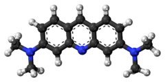
Acridine orange
 | |
 | |
| Names | |
|---|---|
| Preferred IUPAC name
N,N,N',N'-Tetramethylacridine-3,6-diamine | |
| Systematic IUPAC name
3-N,3-N,6-N,6-N-Tetramethylacridine-3,6-diamine | |
| Other names
3,6-Acridinediamine Acridine Orange Base | |
| Identifiers | |
| 494-38-2 | |
| 3D model (Jmol) | Interactive image |
| ChEBI | CHEBI:234241 |
| ChEMBL | ChEMBL81880 |
| ChemSpider | 56136 |
| ECHA InfoCard | 100.122.153 |
| EC Number | 200-614-0 |
| KEGG | C19315 |
| MeSH | Acridine+orange |
| PubChem | 62344 |
| RTECS number | AR7601000 |
| |
| |
| Properties | |
| C17H19N3 | |
| Molar mass | 265.36 g·mol−1 |
| Appearance | Orange powder |
| Hazards | |
| EU classification (DSD) |
|
| S-phrases | S26 S28 S37 S45 |
| NFPA 704 | |
| Except where otherwise noted, data are given for materials in their standard state (at 25 °C [77 °F], 100 kPa). | |
| | |
| Infobox references | |
Acridine orange is an organic compound. It is used as a nucleic acid-selective fluorescent cationic dye useful for cell cycle determination. Being cell-permeable, it interacts with DNA and RNA by intercalation or electrostatic attractions respectively. When bound to DNA, it is very similar spectrally to fluorescein, with an excitation maximum at 502 nm and an emission maximum at 525 nm (green). When it associates with RNA, the excitation maximum shifts to 460 nm (blue) and the emission maximum shifts to 650 nm (red). Acridine orange will also enter acidic compartments such as lysosomes and become protonated and sequestered. In these low pH conditions, the dye will emit orange light when excited by blue light. Thus, acridine orange can be used to identify engulfed apoptotic cells, because it will fluoresce upon engulfment. The dye is often used in epifluorescence microscopy.
Optical properties
At a low pH (3.5), when acridine orange is excited by blue light, it can differentially stain human cells green while staining prokaryotes bright orange for detection with a fluorescence microscope. This differential staining capability allows more rapid scanning of smears at a lower magnification (400x), than by Gram stain (1000x). Bright orange organisms are easily detected against a black to faint green background.
When an acridine orange binds with DNA, it exhibits an excitation maximum at 502 nm (cyan) and an emission maximum at 525 nm (green). When it binds with RNA, the excitation maximum is located at 460 nm (blue) and the emission maximum is located at 650 nm (red). This is all due to the electrostatic interactions occurring when the acridine molecule intercalates between the nucleic acid base pairs.
Acridine orange binding with the nucleic acid occurs in both living and dead bacteria, also other microorganisms. Acridine orange is useful for enumerating the microbes in a sample.
Preparation
Acridine dyes are prepared via the condensation of 1,3-diaminobenzene with suitable benzaldehydes. Acridine orange is derived from dimethylaminobenzaldehyde and N,N-dimethyl-1,3-diaminobenzene.[1]
History
In 1942, Hilbrich and Strugger were first described using acridine orange to detect the fluorchromatic staining of microorganisms. Since then the use of acridine orange has been performed frequently in the examination of soil and water for microbial content. Direct counts of aquatic bacteria by using epifluorescent methods were evaluated by Jones and Simon in 1975. They also determined that the best estimation of the bacterial population in lake, river, and seawater samples can be achieved using acridine orange.
Acridine orange direct count (AODC) methodology has been used in the enumeration of landfill bacteria. A study shows that the use of AODC in marine bacterial populations can be compared favorably to fluorescent oligonucleotide direct counting (FODC) procedures. Direct epifluoresent filter technique (DEFT) using acridine orange is specified in methods for the microbial examination of food and water.
The use of acridine orange in clinical applications has become widely accepted; mainly focusing on the use in highlighting bacteria in blood cultures. In 1980, a study involved the comparing acridine orange staining with blind subcultures for the detection of positive blood cultures was done by McCarthy and Senne. The results showed that the acridine orange is a simple, inexpensive, rapid staining procedure that appeared to be more sensitive than the Gram stain for detecting microorganism in clinical materials. Later on, Lauer, Reller and Mirret performed a similar study, compared acridine orange with the Gram stain for detecting the microorganisms in cerebrospinal fluid and other clinical materials. As a result, they reached the same conclusion that was reported by McCarthy and Senne.
Uses
Acridine orange has been widely accepted and used in many different areas, such as epifluorescence microscopy, the assessment of sperm chromatin quality. Acridine orange stain is particularly useful in the rapid screening of normally sterile specimens, and its recommended for the use of fluorescent microscopic detection of microorganisms in direct smears prepared from clinical and non-clinical materials. The staining has to be performed at an acid pH in order to obtain this differential staining effect with bacteria showing orange stain and tissue components yellow to green.[2]
Acridine orange is a versatile fluorescence dye used to stain acidic vacuoles (lysosomes, endosomes, and autophagosomes), RNA, and DNA in living cells. This method is a cheap and easy way to study lysosomal vacuolation, autophagy, and apoptosis. Acridine orange emission changes from yellow, to orange, to red fluorescence as the pH drops in acidic vacuole of the living cell. Acridine orange emits yellow fluorescence when it binds RNA and green fluorescence when it binds DNA. Nuclei emit yellowish-green fluorescence in normal condition, and deep green fluorescence when RNA synthesis is inhibited by compounds such as chloroquine.[3]
Acridine orange can be used in conjunction with ethidium bromide to differentiate between viable, apoptotic and necrotic cells. Additionally, acridine orange may be used on blood samples to cause bacterial DNA to fluoresce, aiding in clinical diagnosis of bacterial infection once serum and debris have been filtered.[4]
References
- ↑ Thomas Gessner and Udo Mayer "Triarylmethane and Diarylmethane Dyes" in Ullmann's Encyclopedia of Industrial Chemistry 2002, Wiley-VCH, Weinheim.doi:10.1002/14356007.a27_179
- ↑ http://ki.se/sites/default/files/goran_kronvall_ao_staining_mini-review.pdf
- ↑ Chloroquine inhibits cell growth and induces cell death in A549 lung cancer cells. Bioorganic & Medicinal Chemistry. 2006 May 1; 14(9):3218-3222.
- ↑ Acridine Orange Stain. Infection Control.
